Cancer Diagnostic Imaging

Computed tomography (CT) is a diagnostic procedure that uses special x-ray equipment to obtain cross-sectional pictures of the body. The CT computer displays these pictures as detailed images of organs, bones, and other tissues. This procedure is also called CT scanning, computerized tomography, or computerized axial tomography (CAT).
How is CT used in cancer?
Computed tomography is used in several ways:
- To detect or confirm the presence of a tumor;
- To provide information about the size and location of the tumor and whether it the cancer has spread;
- To guide a biopsy (the removal of cells or tissues for examination under a microscope);
- To help plan cancer treatments, such as radiation therapy or surgery; and
- To determine whether the cancer is responding to treatment.
What is PET?
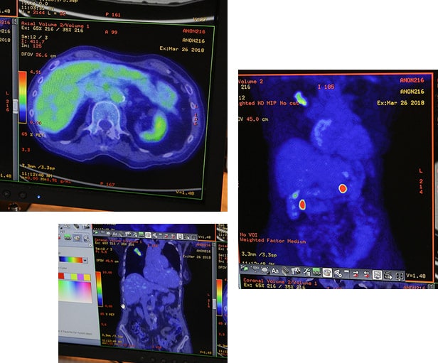
- Positron Emission Tomography (PET) is an advanced imaging technique that produces images of the body’s biological functions.
- Unlike X-rays, CTs (computed tomography), ultrasounds and MRIs (magnetic resonance imaging), PET does not show body structure (anatomy). Instead, it reveals the chemical function (metabolism) of an organ or tissue.
Why is it used?
- PET is used to diagnose and guide the treatment of a number of diseases including cancer, coronary heart disease, neurological disorders and seizure disorders.
- Specifically, PET is a valuable tool for the early detection and diagnosis of lung cancer, breast cancer, melanoma skin cancer, lymphoma, colon and rectal cancers, head and neck cancers, ovarian cancer, pancreatic cancer, and esophageal cancer.
- It is also used for staging and restaging.
- PET may reduce the need for further testing and diagnostic surgeries.
How is a PET scan planned and delivered?
- A PET scan is painless and can be performed in just a few hours as an outpatient procedure. The time required varies depending on what type of scan is being performed, but in most cases, a torso scan from eyes to mid-thigh takes about 2 hours. Some exams, such as brain or heart procedures, take as little as 60 minutes to complete.
- When a patient arrives for a PET scan, he or she will be registered by office personnel and taken
- to the PET area where a technologist will ask a series of medical history questions.
- The patient is then injected with a small amount of radioactive material through an intravenous (IV) line. This substance is called a “tracer” and will be distributed throughout the patient’s body.
- After injection, the patient relaxes and remains relatively still for about an hour.
- For the scan, the patient lies on a “scanning bed” that moves slowly through the PET scanner while the machine detects the injected tracer. When the imaging procedure is complete, the scanner sends the resulting information to a computer that generates several images which are reviewed by a specially-trained physician. A report and pictures detailing the findings are provided to the patient’s physician, usually within a day.
PET Prep Instructions
- Amyvid
- Amyloid
- Cerrianna
- Detectnet
- Illucix
- Locametz
- Netspot
- Neuraceq
- Pluvicto
- Posluma
- Pylarify
- Tau
- Xofigo
Region’s Most Advanced Mobile PET/CT Technology
Together with our fixed 5-ring PET/CT (at NYOH/Albany Medical Center), we offer the region's most advanced PET imaging technology for cancer patients. Our mobile unit offers both PET and CT scanning, allowing a “one-stop” imaging appointment for patients who require both.

Improved Accuracy of Smaller Lesions
General Electric's "Q.Clear" technology helps physicians better detect and define smaller lesions. Using an enhancing algorithm, Q.Clear delivers up to 2X improvement in image quality and accuracy for treatment planning.
Half the Scan Time & Half the Radiation Dose
Our PET cuts scan time in half, using 50% less radiation dose. Regular scans take just 12 minutes (compared to 30 min.). Full body scans finish in 25 minutes (compared to 50 min.) This helps elderly and ill patients, who find it uncomfortable to stay still for longer scanning times.

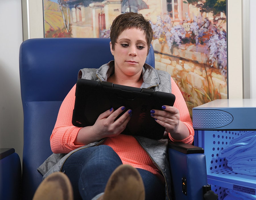
Heated, Reclining Seats, Heated Blankets & iPads
Designed with patient comfort in mind, our custom coach offers fully reclining seats and complimentary iPad use. Heated seats and blankets ensure better scan results.
What Our Patients Say:
Randy Gifford
Colorectal Cancer Survivor, Clifton Park
"You can tell right away that NYOH listened to patients in designing this mobile unit. There is more room, the seats recline and it is brighter inside. Plus, my scan was done much faster. I appreciate having this level of technology just seven minutes from my home. It’s easy to schedule a scan when I need one."
